路阳激发光源用于杨树基因的研究
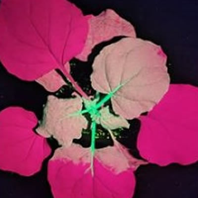
近日,南京林业大学在《Pakistan Journal of Botany》发表文献《OPTIMIZATION OF HAIRY ROOT TRANSFORMATION SYSTEM FOR GENE FUNCTIONAL STUDIES IN POPLAR (POPULUS SPECIES)》,文献中使用了LUYOR-3415RG便携式荧光蛋白激发光源用于观察绿色荧光蛋白GFP在杨树毛状根的表达。LUYOR-3415RG便携式激发光源内置了450nm和530nm两个波段,能够观察绿色荧光蛋白(GFP,eGFP)和红色荧光蛋白RFP(mcherry,tdtomato,dsred2)在动植物上的表达。已经被国内外数千个科学实验室用于基因表达的观察。
文献摘要:
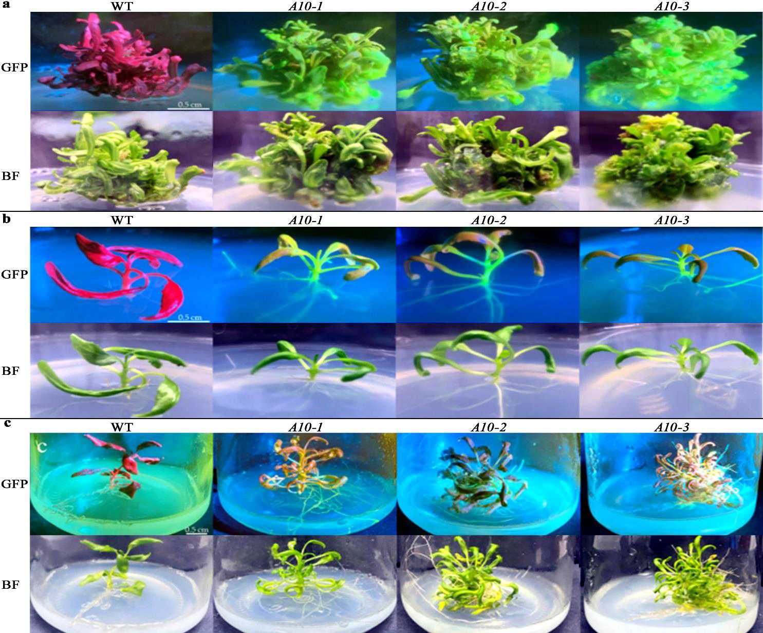
Poplars (Populus spp.) are one of the most widespread ligneous plants in the world, which have a variety of applications ranging from landscape, agriculture, and industry to research. Although Agrobacterium tumefaciens-mediated gene transformation method has been widely utilized for poplar genetic manipulation, many defective technique defects such as the time-consuming procedures, and the occurrence of chimeric transformant have emerged. To address these problems, a simple, fast, straightforward and effective gene transformation method by using Agrobacterium rhizogenes to induce hairy roots in poplars was developed, and regenerated-transgenic poplar plantlets were obtained within a period of two to three months. The optimal procedure comprised the usage of poplar apex buds as explants, infection of A. rhizogenes by directly pricking colonies with the cuttings on the agar plates, and improving hairy-root initiation by application of indole-3-butyric acid (IBA) in combination with hairy-root morphological identification. Moreover, the GFP marker gene was applied to track record the expression level of the introduced genes using a portable fluorescence lamp. Further quantitative real-time PCR (qRT-PCR) and the enzyme activity assay showed that in comparison with the non-transformed control, the transcript abundance and enzyme activity of our target gene (i.e., aspartate aminotransferase) were significantly elevated in the transformed plant lines regenerated from the hairy roots, confirming that the current A. rhizogenes-mediated poplar transformation method is effective and applicable for poplar genetic manipulation and functional genomic study.
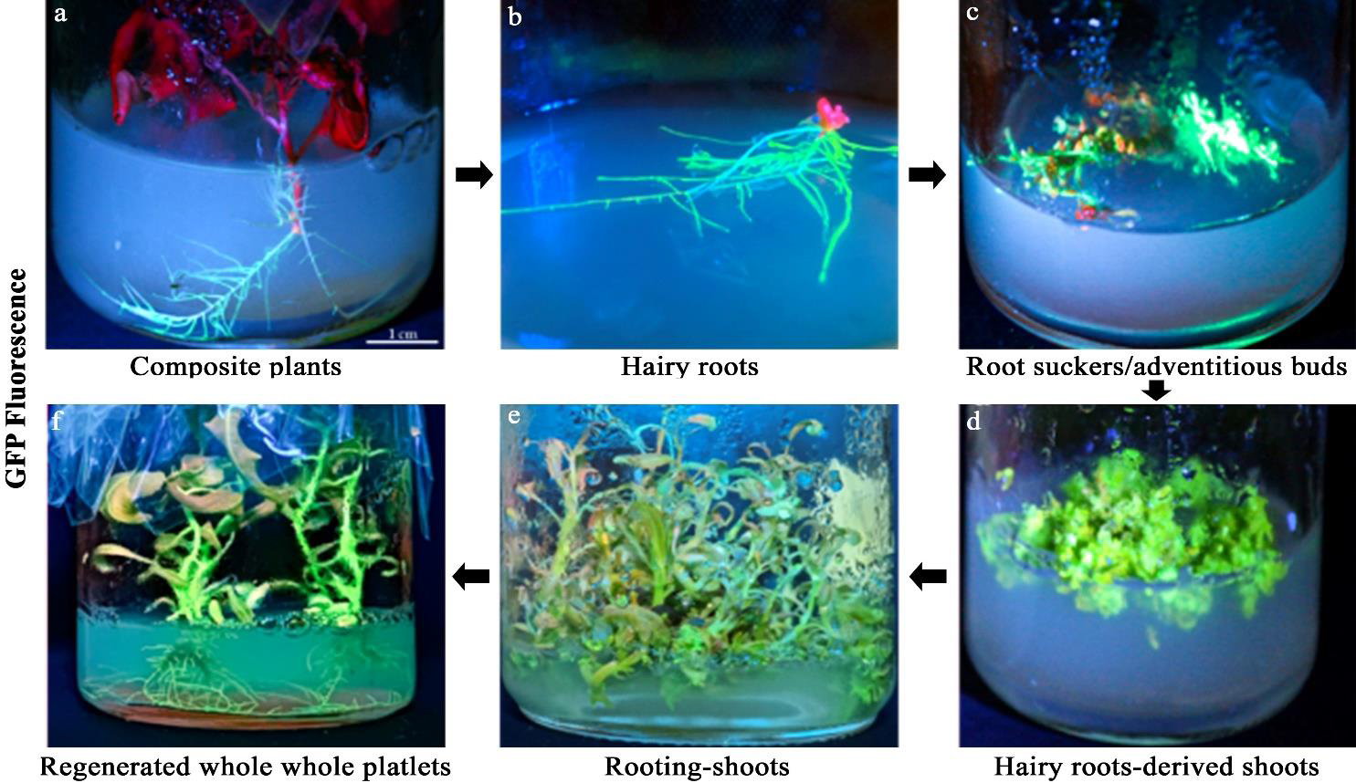
GFP fluorescence detection and the subcellular localization:
The GFP fluorescence was detected under a confocal laser scanning microscope (Carl Zeiss, Jena, Germany). The subcellular localization of the introduced gene was determined by using the 488 nm excitation laser. In addition, the GFP signal for the whole plant was detected by a portable fluorescence lamp (LUYOR-3415, Shanghai, China). The GFP signal (emission at 505-525 nm wavelength), auto-fluorescence (emission at 655–755 nm wavelength) and corresponding bright-field images were photographed at various stages of the transformation procedure.
文献原文下载:
LUYOR-3415RG激发光源产品介绍:LUYOR-3415RG双波长荧光蛋白激发光源
如果您对LUYOR-3415RG激发光源感兴趣,请网站留言或者拨打网页底部联系电话咨询!
 路阳激发光源用于杨树基因的研究
 激发光源用于花青素的生物合成的研究
 高强度紫外线灯用于观察GFP在植物叶片上的表达
 台式紫外灯用于微针制备的固化

